
For nearly 100 years since its discovery, scientists have struggled to fully understand how insulin interacts with its receptor, making it difficult to improve the ability of therapeutic insulins to mimic the way insulin works in the body. Now in a new study, researchers have solved a critical piece of the puzzle by showing how insulin interacts with its receptor at a second binding site. The team of scientists from the Paul Langerhans Institute Dresden (a satellite of Helmholtz Zentrum München) and the Faculty of Medicine Carl Gustav Carus at TU Dresden in Germany, together with colleagues from the Max Planck Institute of Biochemistry in Munich, Germany, McGill University in Canada, and the University of Helsinki in Finland, have published their paradigm-shifting findings in the Journal of Cell Biology.
Over the past century, scientists have detailed the central role of insulin as a regulator of blood sugar and demonstrated its involvement in diabetes and other chronic conditions, including neurodegeneration and cancer. The biological actions of insulin are mediated by its receptor, which is localized to the cell surface. Insulin binds outside the cell on the extracellular domain of its receptor and induces a structural change that is transmitted inside the cell to initiate the response.
“When insulin was administered to patients for the first time in the 1920s, it was a real breakthrough in the treatment of diabetes. However, it is still challenging to generate insulins that recapitulate the full spectrum of endogenous insulin action,” explains Ünal Coskun, group leader at the Institute for Pancreatic Islet Research and Paul Langerhans Institute Dresden (IPI/PLID). “The main reason for this is that we still do not understand enough about how the insulin receptor transmits the signal of insulin binding inside the cell.”
Unlocking the mystery of “site 2”
40 years ago it was established that insulin displays two distinct binding surfaces that interact with the receptor, called “site 1” and “site 2”. While “site 1” interactions with the receptor have been observed, the existence and nature of “site 2” interactions remained, for the most part, a mystery.
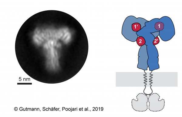
Using cryo-electron microscopy, the researchers were able to demonstrate the 3D structure of the insulin receptor ectodomain, which was saturated by insulin. “The key was to examine the outer part, or ectodomain, of the insulin receptor after saturating it with high concentrations of insulin,” explains Theresia Gutmann, co-first author of the study from the Paul Langerhans Institute and the German Center for Diabetes Research.
Co-first author Ingmar Schäfer, from the Department of Structural Cell Biology at the Max-Planck Institute of Biochemistry (MPIB) adds, “We recorded over 8000 electron microscopic images and analyzed over 300000 single receptor particles, from which we could generate 2D images of the “T”-shaped complex to reconstruct a 3D image.”
The approach allowed the group to capture the binding of insulin to “site 2” for the first time. Since the receptor consists of two identical parts, each containing two insulin binding sites, up to four insulin molecules were bound by the receptor in total (Figure 1).
In parallel, the scientists used computational modelling and simulation approaches to investigate and validate the dynamics of all contact sites with atomistic resolution. “Computational techniques such as these are becoming increasingly important to analyze complicated dynamic processes in living cells, with the added advantage of allowing drug screens to be performed in silico,” says Ilpo Vattulainen from the Department of Physics at the University of Helsinki.
The identified interactions match well with earlier predictions, including those for “site 2,” and the researchers can now reconcile the findings of earlier studies with the structural data they obtained. These newly characterized “site 2” interactions might be important for the initial contact of insulin with the receptor.
The scientists hope that these details concerning insulin–receptor interactions will ultimately expand the current models of insulin binding to its receptor and inform new approaches to structure-based drug design. “Cryo-electron microscopy is a powerful technique, and we are excited to see how these new insulin binding sites will augment the development of designer insulins with more targeted activity,” says co-senior author, Michael Strauss of McGill University’s Department of Anatomy and Cell Biology.
“Cryo-EM structure of the complete and ligand-saturated insulin receptor ectodomain,” by Gutmann, Schäfer, Poojari, Brankatschk, Vattulainen, Strauss, and Coskun, was published in the Journal of Cell Biology on November 14, 2019. DOI: 10.1083/jcb.201907210
November 14, 2019