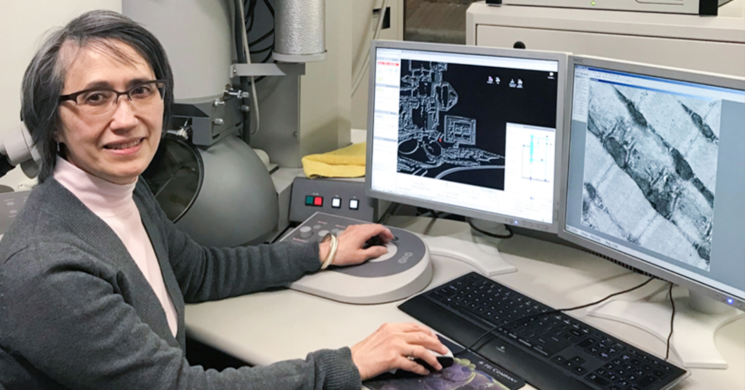
The recently retired Jeannie Mui has been a fixture at McGill’s Facility for Electron Microscopy Research
“I took career aptitude and assessment tests in high school and, based on my interests, skills and personality traits, my guidance counselor recommended a professional or technical career,” shares Jeannie Mui, a native of Hong Kong who emigrated to Canada at the age of 10. “As a first-generation immigrant and the only girl in a family with four older brothers, I thought a professional career was not possible.”
Uncertain about her career path after graduating with a degree in Biology from Concordia University in 1978, a friend of Jeannie’s who was working as a technologist at the Faculty of Engineering at McGill at the time suggested she submit a job application via the University’s central Human Resources department. In August of that year, she received a telephone call from Michael Lalli, PhD, the Director of the Electron Microscopy (EM) Laboratory in the Department of Anatomy & Cell Biology at the time, requesting an interview. Following a final interview with the departmental chair, Yves Clermont, PhD, who, along with Charles P. Leblond, PhD, was a pioneer in biological electron microscopy, Jeannie was hired and began working in the EM Lab in October 1978. “Besides working in a hospital, the next best place for a technical career in science was an academic,” she says.
The importance of lifelong learning
After joining the EM Lab, Jeannie needed to train in various sample preparation techniques appropriate for light and electron microscopy, including animal perfusion, processing of cells and tissues, ultramicrotomy (a method of cutting 70 to 100 µm thick slices or ultrathin sections) and the basic imaging of these ultrathin sections by transmission electron microscopy (TEM). “I also learned to use the darkroom to develop negatives and print micrographs for the Principal Investigators (PI) in the department,” notes Jeannie. “Basic techniques varied on what the PIs wished to image, such as regular ultrastructural studies of morphology or immunolabelling by autoradiography, a technique perfected by Prof. Leblond.”
As she continued to acquire higher technical experience and expertise, Jeannie was promoted to Senior EM Technician in 1981. “I learned to operate a new generation of TEMs, print micrographs for publication and studied new sample preparation techniques, such as slam freezing and automatic freeze substitution, which better preserved biological samples,” she explains. “For example, a graduate student in the department applied the technique of immunogold labelling according to the Tokoyasu method. At the time, no workshops or courses were available for the Tokoyasu method for cryo-ultramicrotomy, so I had to learn the technique by reading the published literature.”
The birth of the FEMR
In 1999, with Hojatollah Vali, PhD as Director and S. Kelly Sears, PhD, as Research Manager, the Facility for Electron Microscopy Research (FEMR) was formally launched. The FEMR grouped many of the EM laboratories at McGill into a single cross-departmental, cross-faculty and cross-institutional core facility.
As Chief EM technician, Jeannie was responsible for the operation of three TEMs, training students in sample preparation and imaging by TEM, and providing technical advice and guidance for research, not only in the life sciences but branching out into the environmental, materials, and physical sciences where Prof. Vali brought his cross-disciplinary expertise to electron microscopy.
“I have worked with Jeannie since joining the Department of Earth & Planetary Science in the fall of 1989,” shares Prof. Valli. “At that time, there were 18 electron microscopes on the downtown campus. However, with respect to the expertise and infrastructure, the EM Lab in Anatomy & Cell Biology was McGill’s only comprehensive EM lab. My research was at the interface of materials and biology and involved investigating colloidal systems and biomineralization. However, Jeannie’s activities were exclusively biological. Still, she never hesitated to take on the complex challenges of adapting her expertise and techniques for biological sample preparation, such as cryo-ultramicrotomy and immunogold labeling, to inorganic systems. Jeannie’s technical knowledge and expertise permitted the research community at McGill to engage in high-quality research collaborations with national and international institutes such as Caltech, MIT, Harvard, UC-Berkeley, and many others. These collaborations often produced high-impact publications in Nature, Science, and PNAS. Without Jeannie’s contributions, none of this research would have occurred. Jeannie is the only expert in Canada capable of working on both biological and non-biological materials.”
As researchers become dependent on advanced instruments, they required highly specialized knowledge and expert training in the proper operation and use of the research infrastructure. “I was also responsible for preparing or embedding and sectioning a new variety of materials, including soft and beam-sensitive materials, clay minerals and other crystalline materials,” says Jeannie. The 2000s also brought a new generation of EMs and imaging modalities. “As I became more involved in research that resulted in publications, I was also responsible for overseeing the work of technical staff and undergraduate and graduate students, and I was promoted to EM Coordinator In 2013.”
Teaching the next generations
“When I train new users in electron microscopy, I always try to place myself in their shoes,” explains Jeannie. “Electron microscopy can be a daunting task; therefore, the aim of my training is primarily an easy-to-follow learning experience with emphasis on hands-on operation rather than on rote memorization. By using everyday scenarios and simple terminology, I try to impart to the students the what, why, how, when, and which techniques to apply for each part of their research project. Thus, the execution of the steps becomes sensibly automatic.” Jeannie notes that confirming that staff is always available for assistance, even after-hours and weekends, reduces damage to instruments through inadvertent misuse or negligence, or mistakes in applying sample preparation protocols. “Since the outcome of any experiment often may depend on my technical work, performance and expertise, my motivation and drive are to use my acquired knowledge and best techniques to ensure the highest quality results to help advance the progression of each research project for the students. It is rewarding to know that some of the grad. students went on to have academic positions in other universities in Canada, US, South America & Europe.”
Jeannie recalls something she learned during her early days of printing 3D stereo photographs as being critical for scientific success. “Three-dimensional stereoscopic photography is the art of capturing and displaying two slightly offset photographs to create a three-dimensional image,” she explains. “Prof. Clermont held up a pair of photographs, took a quick look at the two images without a stereoscope, and immediately told me one of them was not in perfect focus. That’s when I learned that good is not good enough. Every task must be as best as possible. One minor misstep or omission in a long line of procedures may affect a project’s outcome. This is what I have tried to achieve in my 43 years of dedicated service at McGill while working with many world-class researchers and undergraduate and graduate students.”
Recognizing decades of contributions
Last year, Jeannie was the recipient of the Technologist Award from the Microscopical Society of Canada. The award honours a technologist who has made significant contributions to the advancement of microscopy research or training through their role as a technologist at a Canadian university, industry, or government and is one of the most prestigious awards a technologist can receive in Canada.
“I have to admit I was not surprised at all when news from the Microscopical Society of Canada came in about the Technologist Award, “says Joaquin Ortega, PhD, the current FEMR Director. “I knew Jeannie could win this award hands down! There are only a handful of people in North America with Jeannie’s trajectory, dedication, and unique expertise. McGill and FEMR are tremendously fortunate to have Jeannie working with us.”
“I have always enjoyed working in science,” notes Jeannie. “It is the self-satisfaction that drove me to try to keep getting better in the field. I never thought of myself as a technologist. However, I was pleased the Awards Committee of the Microscopical Society of Canada recognized my dedicated commitment to advancing the development of EM techniques and transferring this knowledge to future researchers at the FEMR, McGill University, and beyond. This was reflected in the innumerable numbers of high-quality personnel that I had the opportunity to train in all aspects of electron microscopy and the hundreds or more publications, many in high-impact journals in life sciences, physical sciences, and engineering, where I contributed my technical expertise.”
Although Jeannie retired in October 2021, she continues to work at the FEMR as a casual employee on special research projects, continuing to share the unique expertise she has developed over the years.